The DW-P5 portable ultrasound machine is engineered for functionality, flexibility, and ease of use in a variety of clinical settings. Designed with a sleek, compact body, it’s ideal for both stationary and mobile medical environments.
Featuring dual probe ports, the DW-P5 allows users to switch seamlessly between probes during examinations – improving workflow and diagnostic efficiency. Its high-definition (HD) display delivers crystal-clear, detailed imaging, enabling accurate diagnosis across a wide range of applications.
For added convenience, the DW-P50 offers an optional trolley, making it highly adaptable for point-of-care, outpatient, or bedside use.
Versatile ultrasound for every clinical setting
Advanced 4D Portable Ultrasound
4D imaging
15 inch screen
128GB storage
2 probe interfaces
4D imaging
15 inch screen
128GB storage
2 probe interfaces
Features
Features
Thin and Flexible Body
15" full view medical HD display with an all-in-one keyboard for easy operation.
Multiple Probes Selection
Multiple Probes Selection
Previous
Next
Previous
Next
Exceptional Imaging Processing Functions
Exceptional Imaging Processing Functions
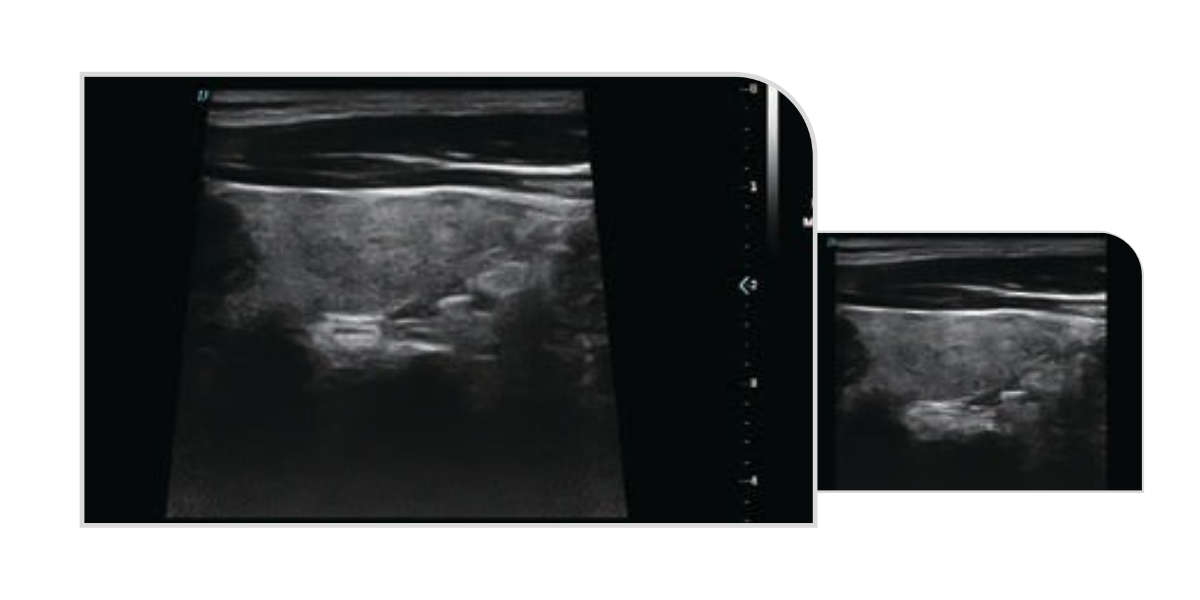
Trapezoidal Imaging
Trapezoidal imaging is an advanced ultrasound imaging technique that expands the standard rectangular view into a trapezoid shape. By widening the left and right sides of the image, it provides a broader field of view, allowing clinicians to visualise more anatomical structures in a single scan - ideal for improved diagnostic accuracy in musculoskeletal, obstetric, and abdominal imaging.
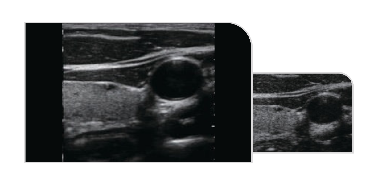
Spatial Composite Imaging
Spatial compound imaging in ultrasound enhances overall image quality by improving contrast resolution, fine resolution, and spatial resolution. It strengthens echo continuity at tissue and lesion interfaces while significantly reducing common artefacts such as speckle, scatter, specular reflection, attenuation, and poor contrast - resulting in clearer, more accurate diagnostic images.
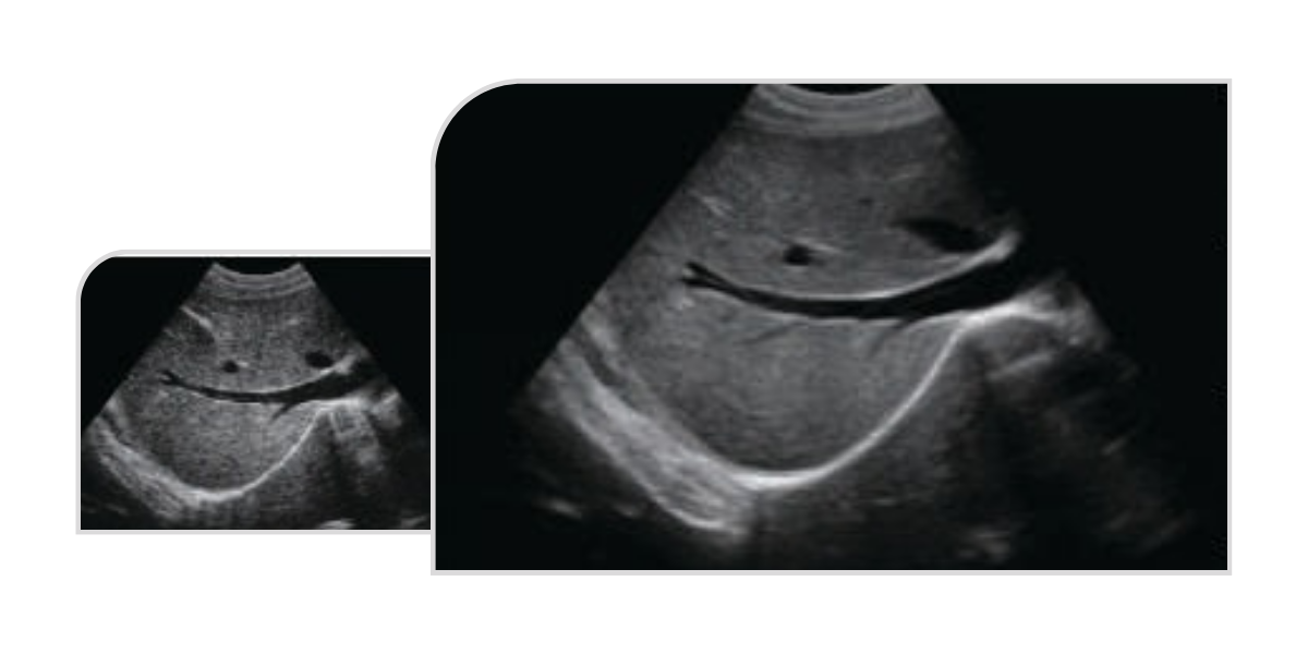
Tissue Harmonic Imaging (THI)
THI enhances image clarity by improving tissue contrast resolution, spatial resolution, and by eliminating near-field artefacts. It is especially useful in the diagnosis of cardiovascular and abdominal conditions, providing clearer visualisation of lesions and more accurate delineation of boundaries - particularly in patients who present imaging challenges.
Micron Imaging
Micron imaging technology enables real-time tracking of signal variations at tissue boundaries to enhance edge definition. It simultaneously monitors each pixel, optimising internal tissue signals while seamlessly integrating edge and internal image data. The result is a highly accurate, detailed, and high-contrast 2D image with exceptional clarity.
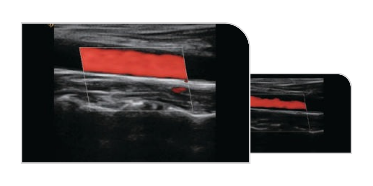
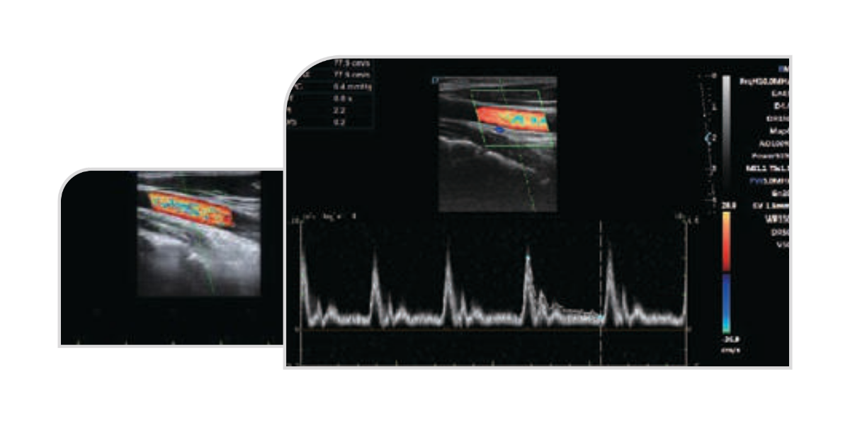
Doppler Spectrum Auto-Envelope
Automatic and precise spectral analysis, providing real-time calculations of key haemodynamic parameters such as blood pressure, heart rate, and resistance index. This feature supports accurate and efficient cardiovascular assessment, enhancing diagnostic confidence and workflow efficiency.
Related Products
Shop for the best selection of healthcare products.