The DW‑T5 is a high-performance colour doppler ultrasound designed to support a broad range of clinical applications. Whether you’re examining abdominal organs, vascular structures, musculoskeletal systems, pediatrics, OB/GYN, or assisting with interventions, the DW‑T5 delivers exceptional image quality, efficient workflows, and intuitive operation to enhance patient care across the board.
Designed for reliability and precision, the DW‑T5 enables healthcare professionals to deliver comprehensive, patient-centric care across multiple specialties.
Engineered for high performance.
Advanced 4D Cart-Type Ultrasound
4D imaging
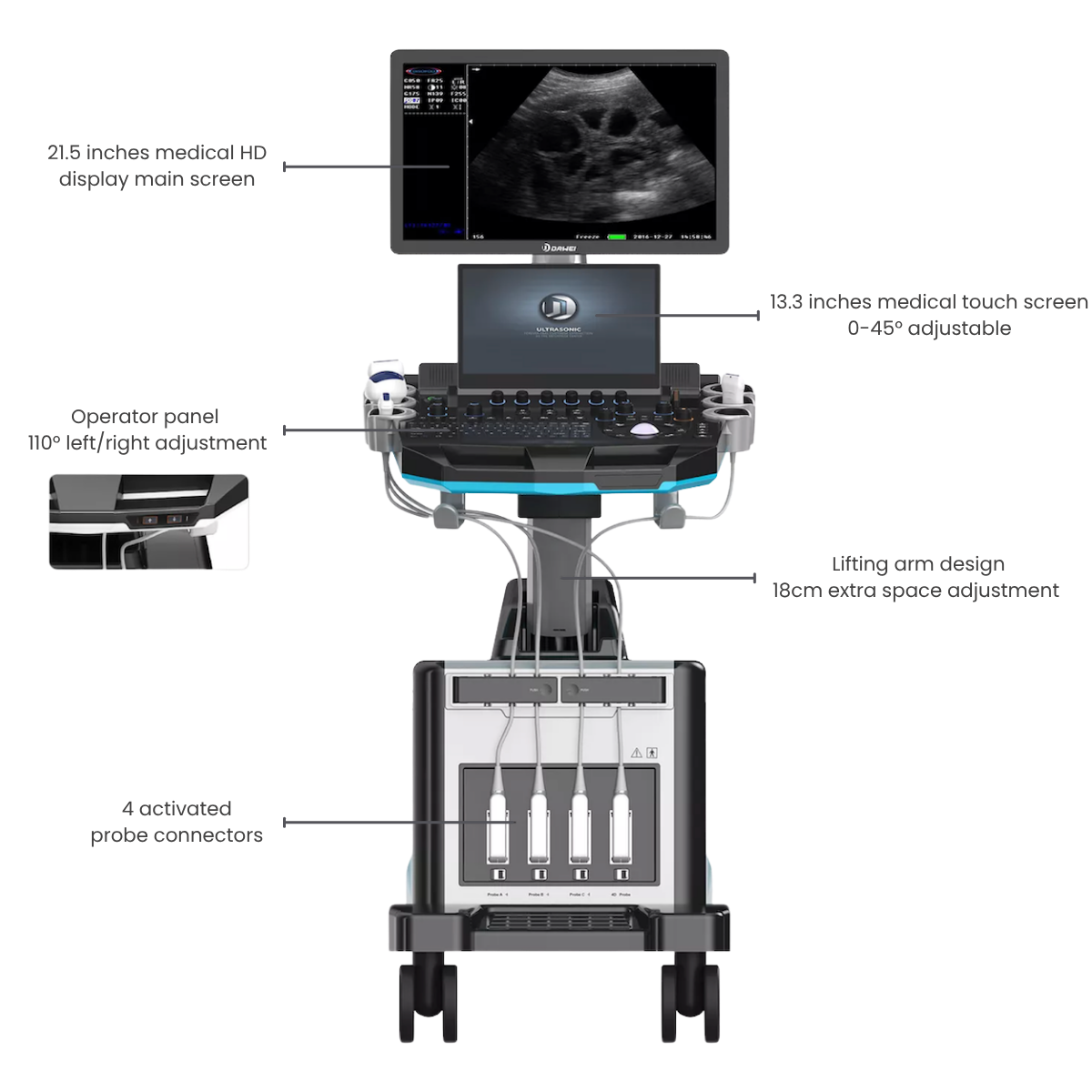
21.5 inch screen
256GB storage
4 probe interfaces
4D imaging
21.5 inch screen
256GB storage
4 probe interfaces
Features
Multiple Probes Selection
Multiple Probes Selection
Previous
Next
Previous
Next
Exceptional Imaging Processing Functions
Exceptional Imaging Processing Functions
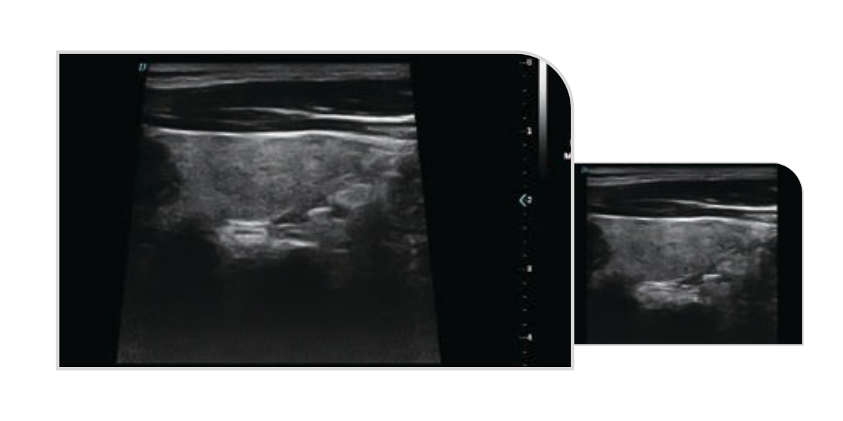
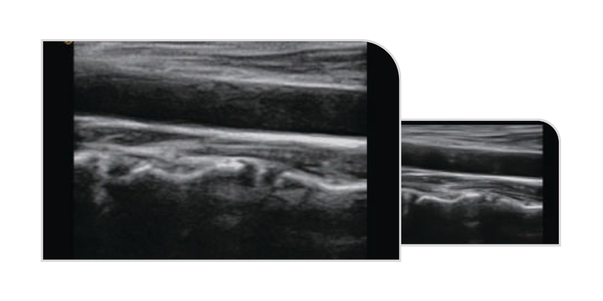
Trapezoidal Imaging
Trapezoidal imaging is an advanced ultrasound imaging technique that expands the standard rectangular view into a trapezoid shape. By widening the left and right sides of the image, it provides a broader field of view, allowing clinicians to visualize more anatomical structures in a single scan - ideal for improved diagnostic accuracy in musculoskeletal, obstetric, and abdominal imaging.
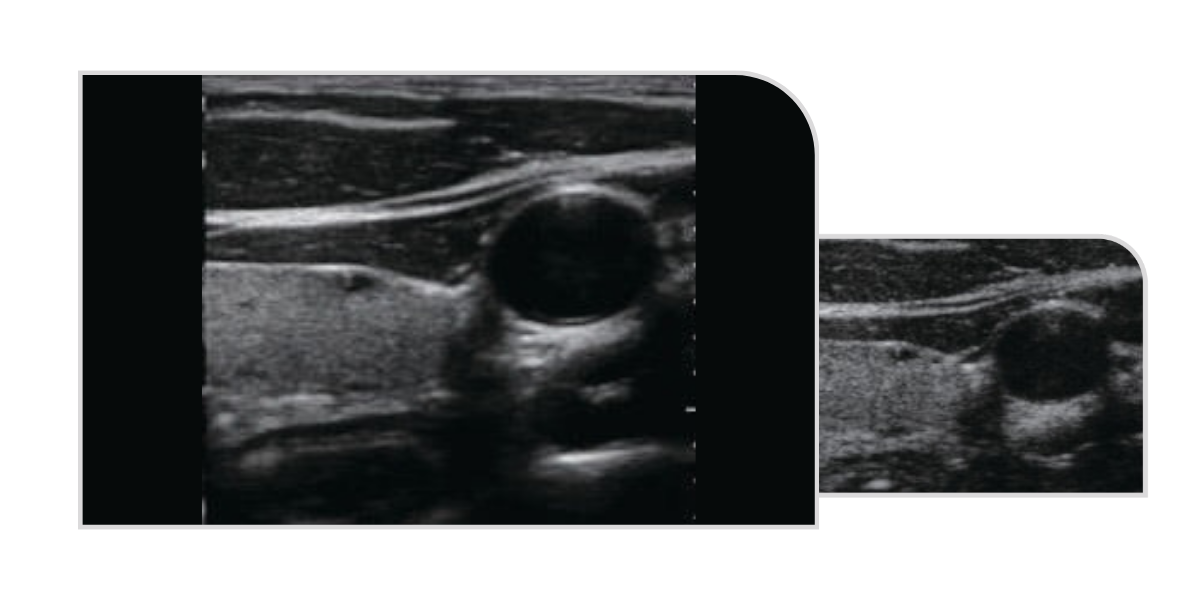
Spatial Composite Imaging
Spatial compound imaging in ultrasound enhances overall image quality by improving contrast resolution, fine resolution, and spatial resolution. It strengthens echo continuity at tissue and lesion interfaces while significantly reducing common artefacts such as speckle, scatter, specular reflection, attenuation, and poor contrast - resulting in clearer, more accurate diagnostic images.
Tissue Harmonic Imaging (THI)
THI enhances image clarity by improving tissue contrast resolution, spatial resolution, and by eliminating near-field artefacts. It is especially useful in the diagnosis of cardiovascular and abdominal conditions, providing clearer visualisation of lesions and more accurate delineation of boundaries - particularly in patients who present imaging challenges.
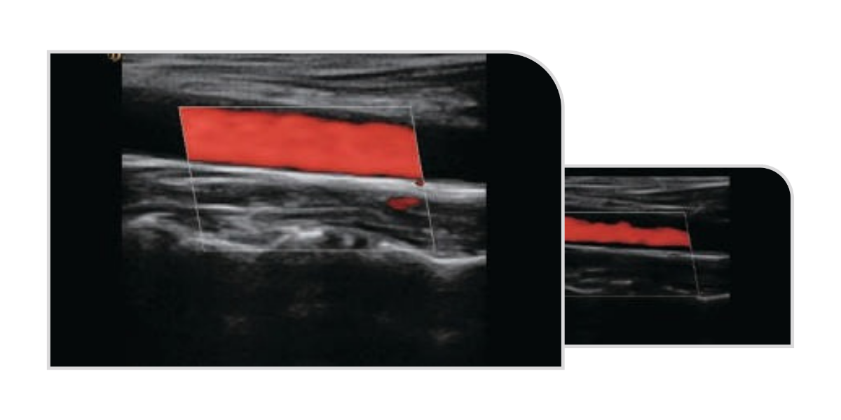
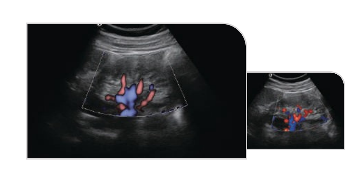
Directional Power Doppler Imaging
DPDI utilises a high-resolution, small sample volume to produce detailed images with dual-colour blood flow direction. This technique minimises colour blooming, offering a more accurate visualisation of vascular structures and defect sizes. Unlike conventional power doppler, it provides clear directional flow information, enhancing diagnostic accuracy in vascular and obstetric/gynaecological imaging.
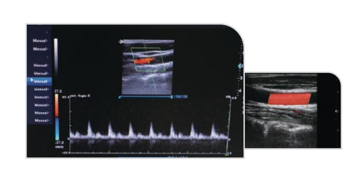
Triple Imaging
Triplex Imaging combines 2D ultrasound, colour doppler, and spectral (pulse wave) doppler for a comprehensive assessment of blood flow. The spectral doppler accurately measures blood flow velocity, making it especially useful in vascular studies. It is commonly used in arterial and venous scans, such as to detect or rule out deep vein thrombosis (DVT) or during carotid ultrasound to assess circulation and identify blockages.
Clean Filter
Clean Filter Technology filters and extracts useful signal information across the full frequency range and at various depths. It analyses signal variation during propagation, applies targeted correction and matching, and effectively suppresses noise. The result is clearer, high-resolution images with improved tissue detail and signal accuracy.
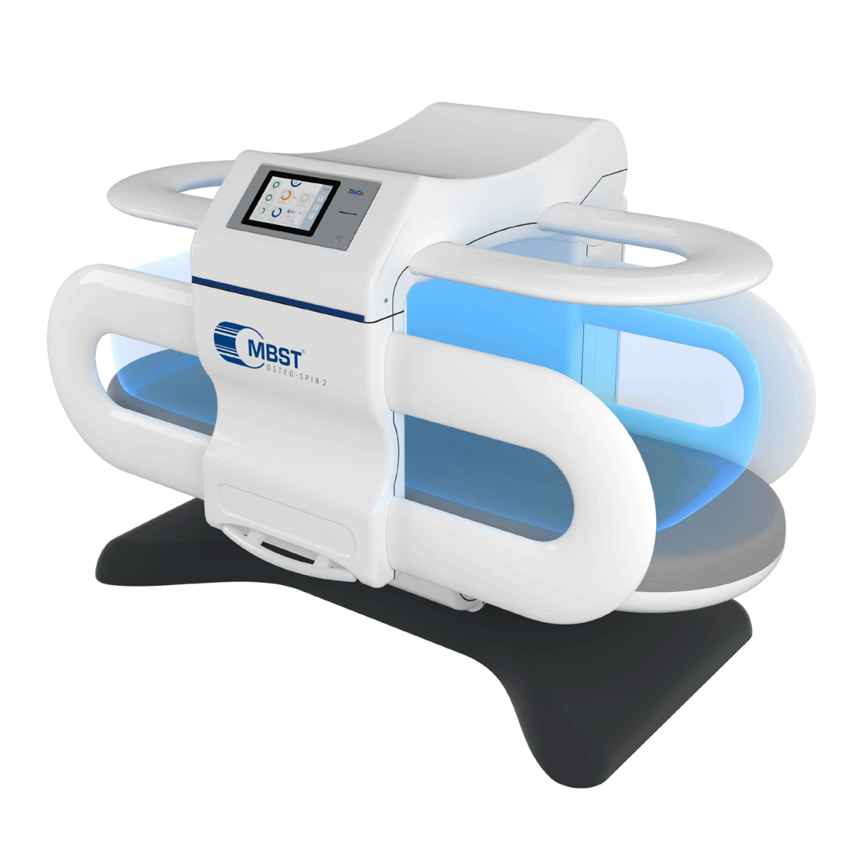
Related Products
Shop for the best selection of healthcare products.